Advanced imaging enabled by 30,000 miniaturized lenses
In many breast and prostate surgeries, surgeons must make critical decisions before definitive pathology results are available. Tissue is removed while the patient is still on the operating table, yet confirmation of whether all cancerous cells are gone often follows hours or even days later. If cancer cells are detected afterward, a second operation or additional treatments like radiation or hormone therapy may be required, adding emotional strain for patients and increasing pressure on healthcare systems. The Histolog® Scanner, developed by SamanTree Medical with support from Sioux Technologies, is designed to close this gap. This medical imaging device enables high-resolution imaging of freshly excised tissue surfaces in one minute or less, while the patient is still on the operating table.

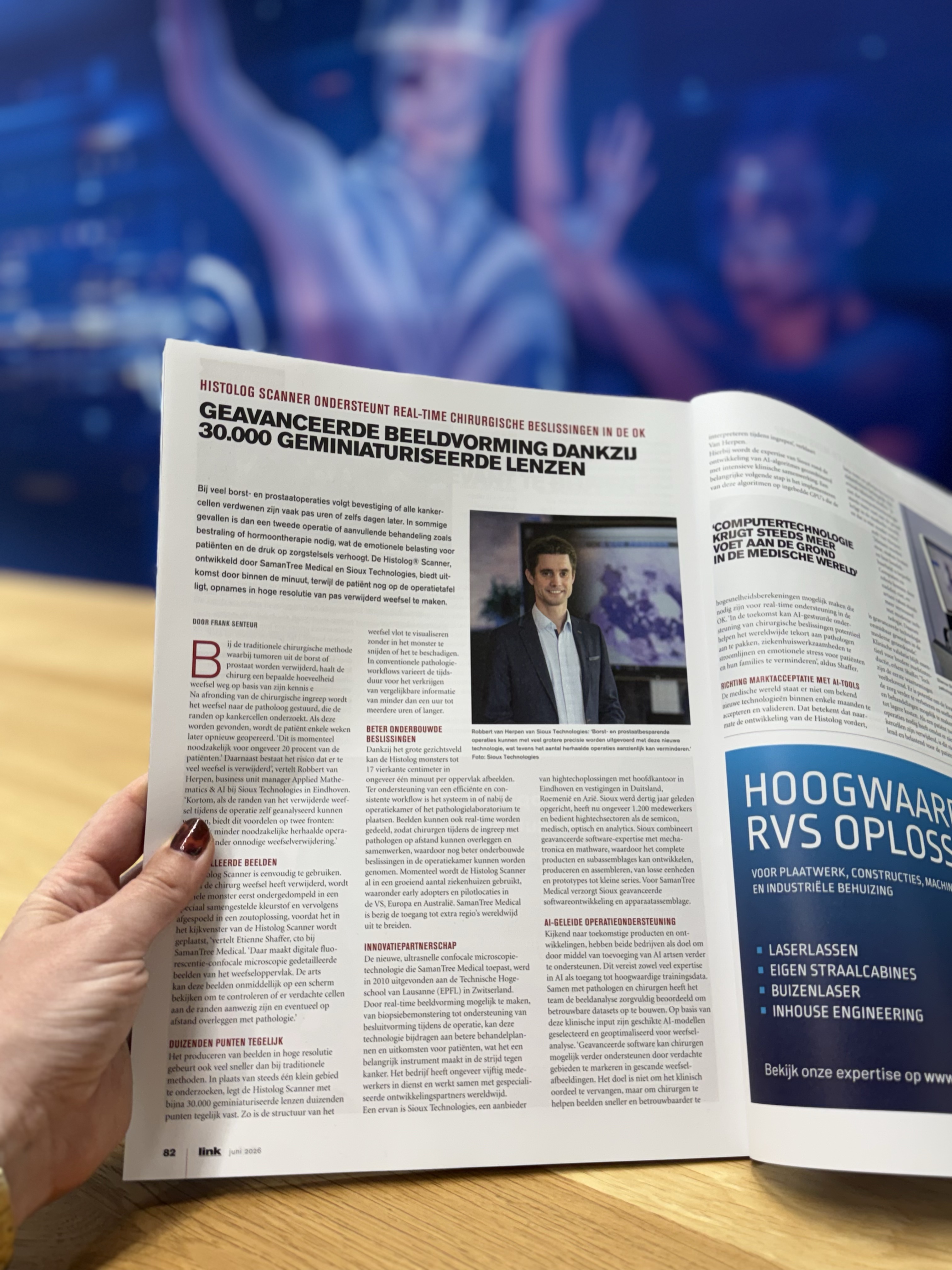
“In the traditional surgical method of removing tumours from the breast or prostate, the surgeon removes a certain amount of tissue based on his knowledge and experience,” says Robbert van Herpen, Business Unit Manager Applied Mathematics & AI at Sioux Technologies in Eindhoven. “After finalizing the surgical procedure, the tissue is sent to the pathologist, who examines the margins for cancer cells. If these are found, the patient is operated on again a few weeks later. This is currently necessary for approximately 20 percent of patients. Next to that, there is a risk that too much tissue has been removed. In short, if the margins of the excised tissue could be analysed during the operation itself, there would be advantages on two fronts: potentially fewer necessary repeat operations and less unnecessary tissue removal.”
Histolog Scanner: real-time imaging
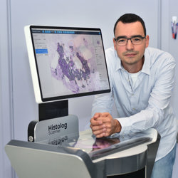
“The Histolog Scanner is easy to use,” says Etienne Shaffer, CTO of SamanTree Medical. “As soon as the surgeon has removed tissue, the entire specimen is first immersed in a specially formulated dye and then rinsed in saline, before being placed on the viewing window of the Histolog Scanner. There, digital fluorescence confocal microscopy captures detailed images of the tissue surface. The physician can immediately review these images on a screen to check whether any suspicious cells are present at the margins or confer with pathology remotely.”
Thousands of points simultaneously
The Histolog scanner produces high-resolution images much faster than traditional methods. Instead of examining one tiny area at a time, it captures thousands of points simultaneously using nearly 30,000 miniaturized lenses. This allows quick visualization of the tissue’s structure without cutting or damaging the specimen. In conventional pathology workflows, obtaining comparable information can take anywhere from under one hour to several hours or longer.
Better-informed decisions
Due to its large field of view, Histolog can image samples of up to 17 cm² in about one minute per surface. The system can be placed in or near the operating theatre or pathology laboratory, supporting an efficient and consistent workflow. Images can also be shared in real time, allowing surgeons to consult and collaborate remotely with pathologists during the procedure, further supporting well-informed decisions in the operating theatre.
Currently, Histolog Scanners are already in use at a growing number of hospitals, including early adopters and pilot sites across the U.S., Europe and Australia. SamanTree Medical is working to extend access to additional regions globally.
Innovation partnership
SamanTree Medical emerged from a new, ultra-fast confocal microscopy technology invented in 2010 at École Polytechnique Fédérale de Lausanne (EPFL) in Switzerland. Their breakthrough enables surgeons to examine tissue margins in high resolution, in real time. By making imaging available for all types of tissue, from biopsy through to physician decision-making during surgery, the technology supports better-informed treatment choices and potentially improved patient outcomes. By enabling real-time imaging from biopsy sampling to intraoperative decision-making, this technology can contribute to better treatment plans and outcomes for patients, making it a crucial tool in the fight against cancer. The company employs approximately 50 people and works with specialized development partners worldwide.
One of its partners is Sioux Technologies, a high-tech solutions provider, headquartered in Eindhoven with offices in Germany, Romania and Asia. Founded 30 years ago, Sioux now has about 1,400 employees and serves the high-tech industry, including semicon, medical, optical and analytics sectors. Sioux combines advanced software expertise with mechatronics and mathware, enabling it to develop, produce and assemble complete products and subassemblies, from single units and prototypes to small series.
Within this partnership, Sioux takes care of the advanced software development and device assembly for SamanTree Medical.
AI-guided surgery assistance
As both companies look to future products and developments, the goal is to add artificial intelligence (AI) support for physicians. Initial work in developing this capability required both strong expertise in AI and access to high-quality training data. Together with pathologists and surgeons, the team carefully reviewed the image analysis to build reliable datasets. Based on this clinical input, suitable AI models were selected and optimized for tissue analysis.
“Looking ahead, advanced software may further support surgeons by highlighting suspicious areas within scanned tissue images. The goal is not to replace clinical judgment, but to help surgeons interpret images more quickly and reliably during procedures,” says Van Herpen.
This work combines Sioux’s expertise in AI algorithm development with intensive clinical collaboration. An important next step is implementing these algorithms on embedded GPUs that enable the high-speed calculations needed for real-time assistance in the operating theatre.
“In the future, AI-guided surgical decision support could help address the global shortage of pathologists, streamline hospital workflows, and reduce emotional stress for patients and their families,” says Shaffer.
Towards market acceptance with AI tools
The medical world is not known for accepting and validating new technologies within a few months. How does this apply to the Histolog Scanner? As the tool moves forward, beta-testing and pilot projects at hospitals will be important, as well as clinical studies.
“You can see that computer technology is gaining an ever-stronger foothold in the medical world, and with good reason,” says Van Herpen. “People get tired, make mistakes and have limited availability. Computers and robots do not have these drawbacks, and doctors are becoming increasingly more comfortable with advanced technology. As such, advanced technology has become indispensable in modern medicine.”
“Clinical validation remains essential for broader market introduction,” says Shaffer. “However, early observations are promising. There is potential to further improve the quality of care and enable faster surgeries and treatments, leading to lower costs. If a patient needs two or more operations because not enough cancer cells were removed, this is not only distressing and burdensome for the patient, but also an extra strain on the healthcare system, both in terms of cost and capacity. The Histolog Scanner illustrates how advanced imaging and software can contribute to this.”